



News
Photobleaching in Fluorescence Imaging: Mechanisms, Impact, and Prevention
Oct 10, 2026
Have you ever noticed fluorescence signals fading during image acquisition, even when your staining appears perfectly fine?
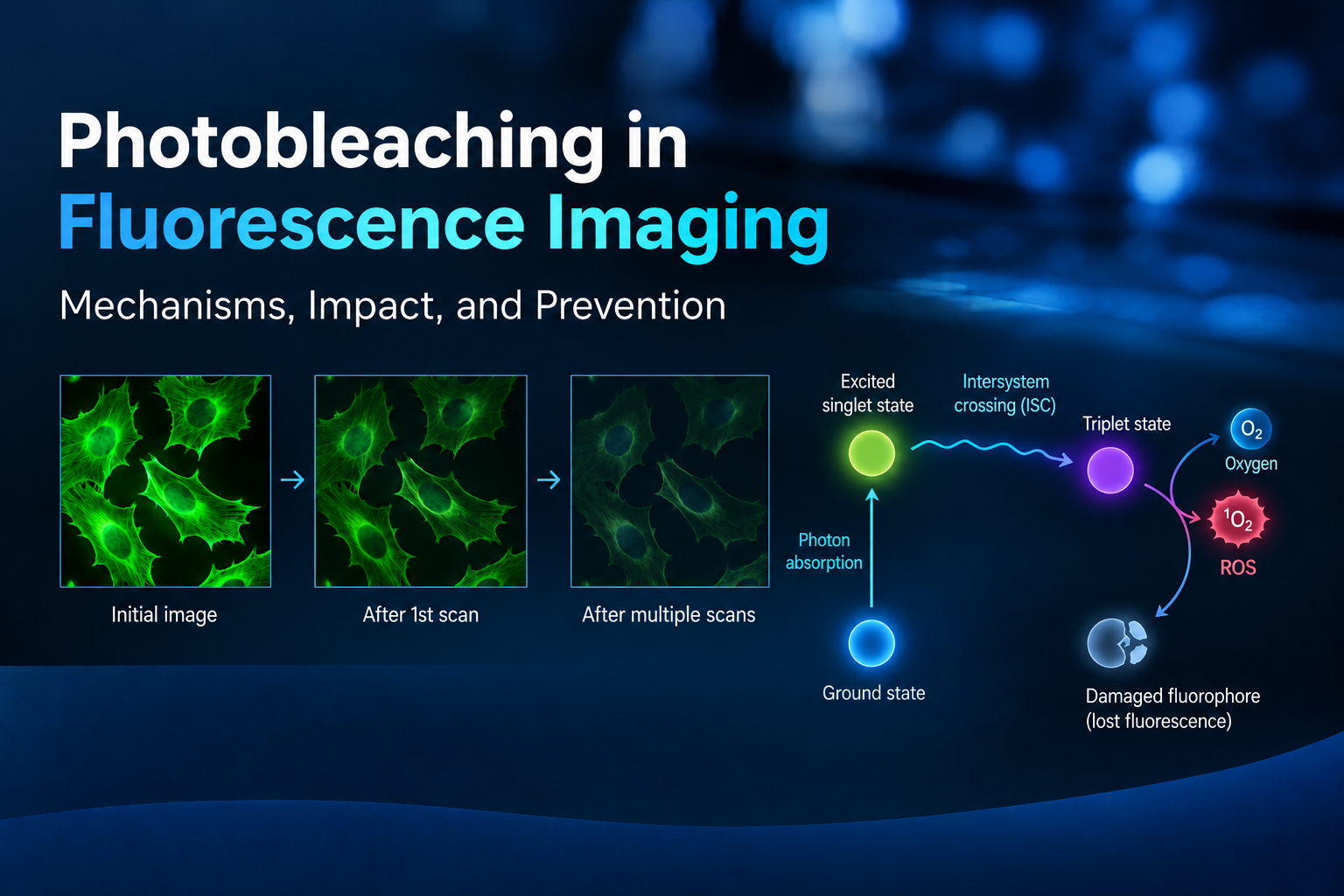
You capture a field of view, adjust the imaging parameters, switch channels, and scan again—only to find that the fluorescence intensity has dropped significantly. After repeated scans, subtle subcellular structures may become difficult to distinguish.
One common culprit is photobleaching, one of the most frequent yet often overlooked challenges in fluorescence microscopy.
What Is Photobleaching?
Photobleaching is the irreversible loss of fluorescence caused by light-induced photochemical damage to fluorescent molecules.
The process typically involves three steps:
• Fluorophores absorb photons and enter an excited singlet state.
• Some molecules undergo intersystem crossing into a longer-lived triplet state.
• Reactions involving molecular oxygen can generate reactive oxygen species (ROS), such as singlet oxygen, which may damage the fluorophore's molecular structure and permanently impair its ability to fluoresce.
Photobleaching depends on fluorophore chemistry, the local environment, excitation intensity, illumination duration, and oxygen availability. In general, stronger or prolonged illumination increases the risk of signal loss.
How to Recognize Photobleaching
Common signs include:
• Fluorescence intensity progressively decreases during repeated imaging of the same region, while the underlying structure remains largely unchanged.
• Weakly labeled subcellular structures become increasingly difficult to resolve.
• In multicolor imaging, one channel loses intensity more rapidly than others.
• Signal loss correlates with imaging exposure rather than simply with sample storage time.
Why Does Photobleaching Matter?
Photobleaching is more than an image-quality issue. It can compromise experimental interpretation and quantitative accuracy.
Quantitative fluorescence analysis: Signal loss can introduce measurement bias.
Live-cell and time-lapse imaging: Fluorescence decay may be mistaken for a biological change.
Weak-target detection: Low-abundance signals can become indistinguishable from background.
Multicolor imaging and colocalization: Unequal bleaching across channels can distort intensity ratios and affect colocalization analysis.
It is also important to distinguish photobleaching from other causes of fluorescence loss. Self-quenching is associated with interactions between nearby fluorophores, while pH-dependent quenching may be reversible. Photobleaching, by contrast, involves irreversible photochemical damage.
How to Minimize Photobleaching
Optimize imaging conditions:
• Use the lowest excitation power that provides an adequate signal-to-noise ratio.
• Minimize exposure time and unnecessary scanning.
• Reduce the number of frames and acquire only essential time points.
• Consider neutral-density filters or more sensitive detectors when appropriate.
Choose photostable fluorophores:
• Consider photostable alternatives, such as Alexa Fluor dyes instead of conventional FITC, when compatible with your application.
• For long-term imaging, evaluate fluorophores under the actual excitation and acquisition conditions.
• In multicolor experiments, account for differences in photobleaching rates across channels.
Optimize the sample environment:
• Use an appropriate antifade mounting medium for fixed samples.
• For live-cell or single-molecule imaging, consider validated oxygen-scavenging or redox-buffering systems when compatible with the biological system.
• Protect samples from unnecessary light exposure and maintain appropriate buffer and mounting conditions.
The key takeaway: Better fluorescence imaging does not always require brighter excitation. Smarter illumination, photostable fluorophores, and an optimized sample environment can help preserve signal quality and improve quantitative reliability.
How do you minimize photobleaching in your fluorescence imaging experiments? Share your experience in the comments!
You capture a field of view, adjust the imaging parameters, switch channels, and scan again—only to find that the fluorescence intensity has dropped significantly. After repeated scans, subtle subcellular structures may become difficult to distinguish.
One common culprit is photobleaching, one of the most frequent yet often overlooked challenges in fluorescence microscopy.
What Is Photobleaching?
Photobleaching is the irreversible loss of fluorescence caused by light-induced photochemical damage to fluorescent molecules.
The process typically involves three steps:
• Fluorophores absorb photons and enter an excited singlet state.
• Some molecules undergo intersystem crossing into a longer-lived triplet state.
• Reactions involving molecular oxygen can generate reactive oxygen species (ROS), such as singlet oxygen, which may damage the fluorophore's molecular structure and permanently impair its ability to fluoresce.
Photobleaching depends on fluorophore chemistry, the local environment, excitation intensity, illumination duration, and oxygen availability. In general, stronger or prolonged illumination increases the risk of signal loss.
How to Recognize Photobleaching
Common signs include:
• Fluorescence intensity progressively decreases during repeated imaging of the same region, while the underlying structure remains largely unchanged.
• Weakly labeled subcellular structures become increasingly difficult to resolve.
• In multicolor imaging, one channel loses intensity more rapidly than others.
• Signal loss correlates with imaging exposure rather than simply with sample storage time.
Why Does Photobleaching Matter?
Photobleaching is more than an image-quality issue. It can compromise experimental interpretation and quantitative accuracy.
Quantitative fluorescence analysis: Signal loss can introduce measurement bias.
Live-cell and time-lapse imaging: Fluorescence decay may be mistaken for a biological change.
Weak-target detection: Low-abundance signals can become indistinguishable from background.
Multicolor imaging and colocalization: Unequal bleaching across channels can distort intensity ratios and affect colocalization analysis.
It is also important to distinguish photobleaching from other causes of fluorescence loss. Self-quenching is associated with interactions between nearby fluorophores, while pH-dependent quenching may be reversible. Photobleaching, by contrast, involves irreversible photochemical damage.
How to Minimize Photobleaching
Optimize imaging conditions:
• Use the lowest excitation power that provides an adequate signal-to-noise ratio.
• Minimize exposure time and unnecessary scanning.
• Reduce the number of frames and acquire only essential time points.
• Consider neutral-density filters or more sensitive detectors when appropriate.
Choose photostable fluorophores:
• Consider photostable alternatives, such as Alexa Fluor dyes instead of conventional FITC, when compatible with your application.
• For long-term imaging, evaluate fluorophores under the actual excitation and acquisition conditions.
• In multicolor experiments, account for differences in photobleaching rates across channels.
Optimize the sample environment:
• Use an appropriate antifade mounting medium for fixed samples.
• For live-cell or single-molecule imaging, consider validated oxygen-scavenging or redox-buffering systems when compatible with the biological system.
• Protect samples from unnecessary light exposure and maintain appropriate buffer and mounting conditions.
The key takeaway: Better fluorescence imaging does not always require brighter excitation. Smarter illumination, photostable fluorophores, and an optimized sample environment can help preserve signal quality and improve quantitative reliability.
How do you minimize photobleaching in your fluorescence imaging experiments? Share your experience in the comments!
Share:


